Looking into the eyes of a cherished dog or cat often provides a clear window into their overall well-being. A pet’s pupils naturally contract and expand in response to changing light levels, expanding in dim environments to gather available light and shrinking in bright sunshine to protect sensitive retinal cells. However, when a companion animal’s pupils remain widely enlarged regardless of ambient light, or when one pupil stays significantly larger than the other, pet owners frequently turn to online searches for trusted vets near me to understand what is happening.
Widely dilated pupils, known medically as mydriasis, can stem from benign behavioral reactions like transient excitement or fear. Yet, persistent or fixed pupil expansion often serves as a key clinical sign of internal ocular pressure shifts, retinal nerve damage, or underlying neurological imbalances. Choosing urgent pet care ensures that veterinary professionals evaluate these delicate optical reactions quickly, providing accurate diagnostic answers and protecting your companion’s vision. For pet parents living in Annapolis, recognizing when dilated pupils reflect a deeper medical concern empowers you to take decisive action before complications arise.
Anatomical Mechanisms Behind Pupil Dilation
To evaluate why pupil responses change, it helps to understand how the iris and nervous system regulate light entering the eye globe.
Autonomic Nervous System Inputs
Pupil size is controlled by the balance between sympathetic and parasympathetic nerve pathways. Stress, excitement, or adrenaline triggers sympathetic nerve activity, causing the iris muscle to pull open. Once the stimulating event passes, parasympathetic signals normally return the pupil to a balanced, responsive state.
Intraocular Fluid Pressure and Glaucoma
When fluid drainage angles inside the eye become obstructed, internal pressure rises rapidly. This mechanical strain forces iris sphincter muscles to fail, leaving the pupil fixed and wide while causing severe ocular pain and corneal fogginess.
Retinal Pathway Disruption and High Blood Pressure
The retina receives light signals and transmits them through optic nerves to the brain. Systemic health shifts, such as elevated arterial blood pressure or internal inflammation, can cause retinal detachment or optic nerve swelling. When light signals fail to reach the brain, the automatic pupil constriction reflex stops functioning.
Evaluating Symptoms: Natural Reactions vs. Medical Concerns
Distinguishing between natural, temporary pupil shifts and a condition requiring intermediate clinical attention depends on observing your pet’s overall posture, lighting environment, and movement.
- Natural Home Variations (Safe for Observation):
- Symmetrical pupil expansion during intense indoor play, hunting toys, or sudden excitement
- Temporary dilation occurring in low-light, dim, or nighttime household settings
- Normal, energetic movement with complete navigational confidence around furniture
- Smooth, equal contraction of both pupils when stepping into bright sunlight
- Clinical Indicators Requiring Urgent Vet Care
- Pupils that remain fully wide and unresponsive when exposed to direct, bright light
- Anisocoria, where one pupil is visibly larger or smaller than the opposite eye
- Unexplained bumping into doorframes, walls, or reluctance to navigate staircases
- Persistent cloudiness, bluish discoloration, or blood spots visible within the eye
- Continuous squinting, pawing at the face, head tilting, or sudden lethargy
Point-of-Care Diagnostics for Pupil Abnormalities
When you seek urgent vet care for abnormal pupil responses, clinical teams rely on swift point-of-care tools to inspect internal eye structures and nervous system function without delay.
- Ophthalmic Tonometry Testing: Measures intraocular fluid pressure using a gentle, soft-tipped probe to screen instantly for pressure spikes associated with acute glaucoma.
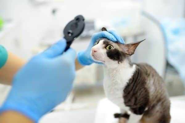
- Direct and Indirect Ophthalmoscopy: Delivers high magnification to inspect lens clarity, examine retinal alignment, and evaluate optic nerve head color and structure.
- Consensual Pupil and Menace Response Tests: Evaluates neurological pathways connecting the eyes to the brain using light reflexes and subtle hand movements.
- Systemic Blood Pressure Screening: Measures arterial blood pressure using an inflatable cuff placed gently around a leg or tail to identify underlying hypertension.
Combining rapid diagnostic findings with pet-safe therapies, anti-hypertensive medications, or pressure-regulating eye drops stops discomfort at its source and preserves vital sight.
Supporting Companion Animals Across Annapolis
Maintaining your pet’s optical health requires attentive daily care, clean surroundings, and staying mindful of subtle changes in their physical appearance. Checking your companion’s eyes during routine grooming sessions or after outdoor exercise allows you to catch minor pupil asymmetries or corneal haze before mobility is affected.
If you are from Annapolis and looking for urgent vet care, taking fast action at the first sign of fixed dilated pupils, cloudiness, or spatial disorientation gives your pet the best opportunity for a smooth, full recovery.
Providing timely medical attention when health changes occur preserves your companion’s quality of life and offers complete peace of mind for your entire household.
Taking Action for Your Pet’s Visual Health
When your dog or cat exhibits persistent dilated pupils, spatial hesitation, or visible physical discomfort, securing a prompt evaluation keeps their recovery moving in the right direction. You can schedule an online appointment through our portal to reserve a convenient time for your pet to be examined by our medical staff.
If your companion is actively displaying uncomfortable eye symptoms or sudden optical changes and you prefer to speak directly with our clinical staff before arriving, please call our team at 410-268-6994 so we can offer immediate guidance and prepare for your visit.
Our dedicated medical team looks forward to delivering gentle, expert care for your companion at Bay Ridge Animal Hospital.
Frequently Asked Questions
Q1. Why are my pet’s pupils dilated even in a brightly lit room?
Ans: When pupils remain wide under bright light, the automatic constriction reflex is impaired. This often indicates retinal damage, optic nerve inflammation, elevated intraocular pressure, or high blood pressure that prevents the eye from processing incoming light normally, requiring prompt veterinary evaluation.
Q2. What does it mean if only one of my pet’s pupils is dilated?
Ans: Unequal pupil sizes, known as anisocoria, suggest an asymmetrical neurological or ocular issue. Common triggers include localized eye trauma, corneal ulcers, unilateral glaucoma, anterior uveitis, or nerve pathway disruptions, all of which warrant swift clinical examination.
Q3. Can stress or fear cause a pet’s pupils to stay dilated for long periods?
Ans: While adrenaline from sudden stress or fear causes temporary pupil dilation, pupils should return to normal size once the pet relaxes in a quiet room. If dilation persists for hours after the stressful event ends, an underlying medical issue rather than emotional stress is likely responsible.
Q4. Are dilated pupils in senior pets always a sign of total vision loss?
Ans: Not necessarily. While dilated pupils can accompany retinal detachment or cataracts in older pets, iris sphincter atrophy (a natural, non-painful weakening of iris muscles in aging animals) can also cause pupils to stay wide without causing complete blindness.
Q5. How should I safely transport a pet with fixed dilated pupils to the clinic?
Ans: Keep your vehicle cool, quiet, and shaded from direct sunlight, as fully dilated pupils allow excessive light into the eye and cause glare sensitivity. Transport small pets in a padded carrier covered loosely with a light sheet, and keep larger dogs calm on a short leash.